
Diagnostic imaging is the noninvasive method of making medical images of your pets to diagnose disease. We have some of the most advanced imaging technologies available for our veterinary patients, of the same caliber found in a modern human hospital.
Our diagnostic imaging capabilities include the full breadth of modalities:
These services are run by our team of on-site and experienced technical staff.
Our state-of-the-art equipment can serve the medical imaging needs of small companion animals (dogs and cats).
Computed Radiography uses a cassette system with an imaging plate that contains photostimulable (light-stimulated) storage phosphors. These phosphors detect and store energy from the x-rays that strike the cassette. When bones and other tissue are placed between the plate and the x-rays, they block the x-rays, allowing an image of those bones to be captured.
When using Computed Radiography, the cassette is run through a computer scanner that uses a scanning laser to release the energy from the plate in the form of light. The light is captured, recorded, and processed into an image by a computer. The imaging plate is then erased by fluorescent light in the scanner, and is ready to be used again.
Ultrasonography is the second most commonly used imaging format in veterinary practice. It uses ultrasonic sound waves in the frequency range of 1.5-15 megahertz (MHz) to create images of body structures based on the pattern of echoes reflected from the tissues and organs being imaged. Several types of image formats can be displayed. The most familiar one (and the one that creates the actual image of anatomy) is B-mode grayscale scanning. The sound beam is produced by a transducer placed in contact with and acoustically coupled by means of a transmission gel to the animal.
An echocardiogram is an ultrasound image of the heart and associated large blood vessels. Echocardiography is a non-invasive test that allows a veterinarian to directly evaluate the anatomy and function of the heart. The examination is performed with the pet lying on a padded table. Most animals do not require sedation (tranquilizers) but some are more relaxed if these are used. A specific form of echocardiography called Doppler allows the cardiologist to look specifically at blood flow which helps to identify leaks in the heart valves, abnormal movement of blood between the left and right sides of the heart, and obstructions to normal blood flow.
Fluoroscopy is a type of advanced imaging that allows us to view the inside of the animal's body while it is in motion. The image is similar to that of a radiograph, or x-ray, but instead of viewing a single still image, many images are produced by fluoroscopy to capture motion in real time. Essentially, this produces an x-ray movie that we can watch and replay. This moving image is helpful to view both the structure and function of organs to identify abnormalities that may not be apparent on a traditional x-ray that shows a single snapshot in time./p>
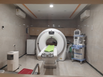
CT (computed tomography, also known as CAT scan) is a diagnostic tool that has long been essential in human medicine and will revolutionize veterinary medicine.
CGS Hospital is delighted to be the first corporate veterinary practice in India to offer CT scanning. We have the only veterinary GE- 16 slice CT scanner in India.
A CT scan is a specialized X-ray test. It can give quite clear pictures of the inside of your body. In particular, it can give good pictures of soft tissues of the body which do not show on ordinary X-ray pictures.